Sponge Choanocyte (PORP_0000003)
A sponge choanocyte, also known as a collar cell, is a specialized cell type with many important functions. Choanocytes play a crucial role in the feeding, respiration, and water circulation processes within a sponge's body. These cells have a characteristic collar-like structure surrounding a single flagellum. The coordinated movement of the flagella generates water currents, drawing water and suspended particles which include food and oxygen into the sponge. The collar captures these particles, allowing the sponge to filter-feed and respirate oxygen. In addition to their feeding function, choanocytes also contribute to the sponge's reproduction through their potential to differentiate into other cell types.
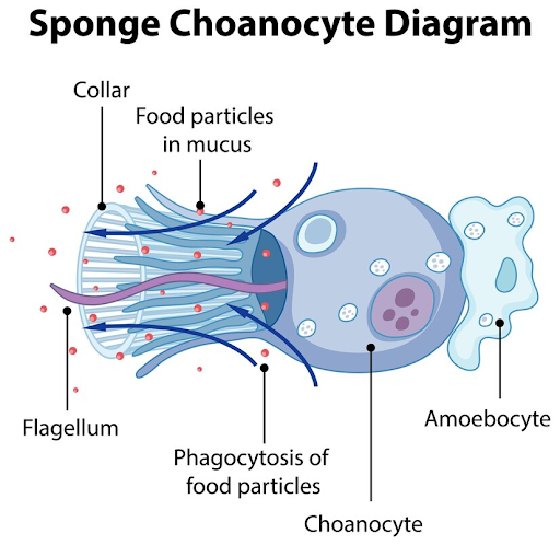
Above is a graphic of Choanocyte with copyright from Freepik.
Note: https://docs.google.com/document/d/1oEJobsgxXRT0IoXLgtnivcRIX0zmyh-mLSSLuMGVUZk/edit.
Cell Ontology (CL) ID: PORO_0000003
CL definition: A cell that lines the interior of Asconoid, syconoid and leuconoid body type sponges that contain a central flagellum surrounded by a collar of microvilli which are connected by a thin membrane. The choanocyte is ovoid, with one end adjacent to the mesohyl (in some species). It is the closest family member to the free-living ancestor called choanoflagellate. The flagellae beat regularly, creating a water flow across the microvilli which can then filter nutrients and other food from the water taken from the collar of the sponge. Food particles are then phagocytosed by the cell. Choanocytes are found dotting the surface of the spongocoel in asconoid sponges and the radial canals in syconoid sponges, but they comprise entirely the chambers in leuconoid sponges. By cooperatively moving their flagella, choanocytes generate a flow of water through the sponges pores, into the spongocoel, and out through the osculum. This improves both respiratory and digestive functions for the sponge, pulling in oxygen and nutrients and allowing a rapid expulsion of carbon dioxide and other waste products. Although all cells in a sponge are capable of living on their own, choanocytes carry out most of the sponge's ingestion, passing digested materials to the amoebocytes for delivery to other cells.
Synonyms: collar cell; gastral cell
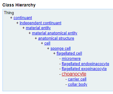
Cell Hierarchy (defined by CL):
Choanocyte hierarchy according to the PORO Ontology:
Above is a graphic of the cell hierarchy of choanocyte and its associated cell types represented in the PORO. Later on we will show a cell hierarchy with live links to other cell types.
Location (defined by CL and UBERON):
The following figure shows the anatomical location of choancytes:
Source of the above figure: https://www.hiclipart.com/free-transparent-background-png-clipart-xrleg/download.
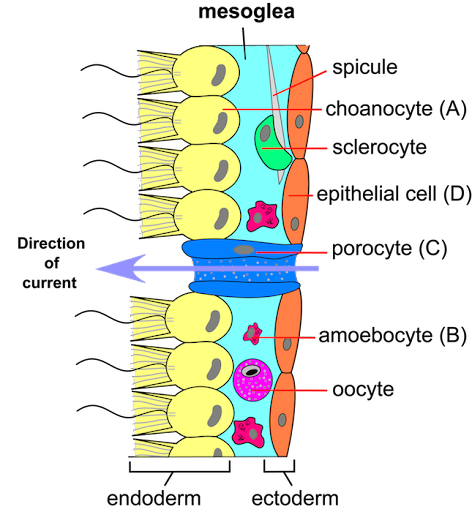
Connections and Vicinity:
Choanocytes are neighbors of many other cell types as shown in the above figure.
Specifically, the neighborhood cell types of Choanocyte include:
- Amoebocyte
- Choanoderm
- Porocyte
- Archeocyte
- Pinacocyte
- Laphocyte
- Sclerocyte
- Oocyte
Cell Differentiation and Lineage:
The following figure shows the cell lineage of choancytes:
Permission of using this figure from Funayama et al granted by the publisher (Wiley.com).
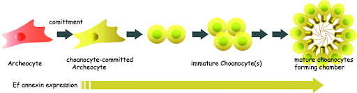
Choanocytes are an essential part of the Amphimedon stem cell system. During metamorphosis, postlarval archeocytes form choanocyte chambers from either a single progenitor cell or multiple cells. Choanocyte chambers grow by proliferation of choanocytes within the chamber and by recruitment of choanocytes into mature chambers. Ref: PMID: 26958337.
Cell Type Evolution Among Organisms:
There are structural similarities between unicellular choanoflagellates and sponge choanocytes demonstrates evolution of multicellularity. Ref: Sogabe et al, 2019.
Cell Biomarkers:
Choanocyte-related biomarkers:
- Ef annexin - Lineage marker: Ef annexin is initially expressed in the choanocyte-committed archeocyte, and then undergoes several mitotic cell divisions to form a choanocyte chamber. Ref: PMID: 15921499.
Gene Expression Profiles:
Specific gene expression profiles are provided below:
- EmH-3: The EmH-3 gene is expressed differentially in the course of development and in a cell-type specific manner. EmH-3 is strongly expressed in the pluripotent archaeocytes, whether isolated from fully differentiated sponges (adult archaeocytes) or from HU-treated sponges (embryonic archaeocytes). Conversely, in differentiated cells such as pinacocytes and choanocytes, EmH-3 expression is very weak and similar to that found in the resting gemmules. Overall, EmH-3 plays a role in cell determination and/or differentiation, and it would determine which archaeocytes will multiply and undergo differentiation and which ones will remain undifferentiated. Ref: PMID: 10576335.
- prox1: prox1 from Ephydatia fluviatilis is another freshwater sponge homeobox-containing gene. It is expressed almost at the same level at all stages of development and in all the investigated cell populations. Ref: PMID: 10576335.
Pathways and Functional Maps:
Podocyte-associated pathways and functional maps:
- Six signaling pathways are conserved across Metazoa: Wnt, TGF-beta, Hedgehog, Notch, FGF and NO-dependent. Ref: PMID: 31036299.
- Wnt signaling is the best studied pathway in sponges due its probable role in axial patterning, especially in early branching Metazoa. The champion in Wnts complexity, a calcisponge Sycon ciliatum, contains 21 ligands. In addition to ligands, four receptors and all core intracellular components except Axin are found in S. ciliatum. Ref: PMID: 31036299.
References:
- All sponges cells in Cellcards: https://cellcards.org/organisms/sponges/.
- Wiki: https://en.wikipedia.org/wiki/Choanocyte.
- Ontobee search for Choanocyte: https://ontobee.org/search?ontology=&keywords=Choanocyte&submit=Search+terms.
Note: This is a manually generated website. Lessons learned will be used for automatic cell card content generation. Feedback is welcome. Thanks!